1. Introduction
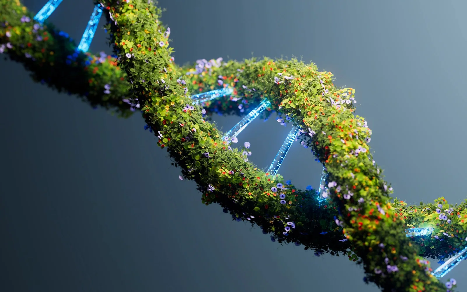
DNA is a helix, not a flat spiral. This distinction is fundamental: a helix is a three-dimensional curve that winds around a cylindrical axis at constant pitch, while a spiral is a flat curve that moves away from a center. The DNA double helix is one of the most important structures in molecular biology.
2. Structure of the Double Helix
The DNA double helix, described by Watson and Crick in 1953 from Rosalind Franklin's X-ray diffraction data, is composed of two antiparallel strands wound around each other. Each strand is a polymer of nucleotides (adenine, thymine, guanine, cytosine) linked by a sugar-phosphate backbone. The two strands are held together by hydrogen bonds between complementary bases (A-T, G-C).
3. Why a Helix?
The helical shape of DNA results from several physicochemical constraints. Base stacking is stabilized by van der Waals interactions and hydrophobic effects: the flat bases stack like coins, and this geometry imposes a rotation of ~36° between successive bases. Hydrogen bonds between complementary strands constrain the interstrand distance. The aqueous environment favors exposure of the charged backbone to water and burial of hydrophobic bases inside.
4. A, B and Z Forms
DNA can adopt several conformations depending on conditions. Form B (Watson-Crick) is the most common in cells: right-handed helix, 10 base pairs per turn, pitch of 3.4 nm. Form A appears under low hydration conditions: wider and shorter helix. Form Z is a rare left-handed helix that appears in certain GC-rich sequences under torsional stress.
5. Other Biological Helices
- Protein α-helix: 3.6 amino acids per turn, stabilized by intramolecular H-bonds
- Collagen triple helix: three intertwined polypeptide chains
- RNA: local helices in secondary structures (stem-loops)
- Microtubules: helical protofilaments forming a hollow cylinder
6. How the structure was established
The double helix did not arise from a direct photograph of the molecule. Oriented DNA fibers produced X-ray diffraction patterns whose cross geometry indicated helical organization. Reflection positions and intensities constrained pitch, diameter, and stacking. Franklin and Gosling distinguished hydrated and less hydrated states, showing that environment changes conformation. [1]
Watson and Crick integrated these constraints with nucleotide chemistry and specific base pairing. Their model placed sugar-phosphate backbones outside and paired bases inside. A–T and G–C complementarity explained both a regular diameter and a possible copying mechanism. It was a testable structural inference, not a visual resemblance. [2]
7. Geometry of B-DNA
In canonical B-DNA, the two antiparallel strands form a right-handed helix. Base pairs stack approximately perpendicular to the axis, with about ten pairs per turn in the classical description and a rise near 0.34 nanometer per pair. The atomic structure of a dodecamer later showed more than a complete turn and revealed local sequence-dependent variation. [3]
The winding produces a major groove and a minor groove. Proteins can read chemical patterns there without fully opening the double helix. Groove width, hydration, and flexibility vary with sequence and ions. A uniform twisted-ladder representation is therefore useful at large scale but hides local deformations used in molecular recognition.
8. Why the helix is stable
Hydrogen bonds contribute to pairing specificity, but they are not the sole stabilizers. Base stacking, exclusion of water from aromatic surfaces, electrostatic interactions, and ions surrounding charged phosphates all contribute to free energy. The observed structure is a thermodynamic compromise depending on solvent, temperature, sequence, and mechanical constraint.
Separating the strands therefore requires disrupting both stacking and pairing. Helicases use chemical energy to move along DNA; polymerases temporarily stabilize geometries suited to copying. A biological helix is not a passive spring: it is continually bent, opened, twisted, and repaired by molecular complexes.
9. A-DNA, B-DNA, and Z-DNA are not decorative variants
A-DNA is a shorter, wider right-handed helix favored by reduced hydration and found in several RNA-containing duplexes. B-DNA dominates under many physiological conditions. Z-DNA is a left-handed double helix whose backbone follows a zigzag path and is favored by particular sequences and ionic conditions.
Crystallography of an alternating CG fragment established the left-handed atomic structure of Z-DNA, with twelve pairs per turn in that crystal. This discovery shows that the same general chemical composition can occupy distinct conformational basins. Chirality is therefore not an arbitrary label: it describes the actual orientation of the molecular screw. [4]
10. Twist, writhe, and topology
In closed circular DNA or a domain whose ends cannot rotate freely, linking number is constrained. It decomposes into local twist of the double helix and writhe of its axis in space. Adding or removing turns can therefore convert twist into supercoiling, like a telephone cord forming loops.
Topoisomerases change linking number by temporarily cutting one or both strands and then resealing them. This is essential for replication and chromosome segregation. It also shows why helix, superhelix, and planar spiral must be distinguished: they belong to different geometric and topological descriptions.
11. What a molecular model must specify
An image of DNA should state at least the represented form, handedness, scale, and level of simplification. A ribbon without atoms describes global architecture; an all-atom model can show pairing but is often still an average or crystal structure. Molecules in solution fluctuate around these conformations rather than remaining frozen.
The strength of the double-helix model comes from convergence among diffraction, chemistry, modeling, atomic structures, and functional experiments. No single source tells the entire story. This methodological convergence is stronger than the cultural icon of the helix, however recognizable it may be.
12. An average structure in a dynamic molecule
At ordinary temperature, base pairs breathe, grooves fluctuate, and the axis bends. Sequences enriched in particular bases may be stiffer, easier to open, or favor a particular curvature. Crystal structures provide extremely useful atomic coordinates, but the crystal, ions, and neighboring contacts select only part of the ensemble of conformations accessible in solution.
Solution experiments, magnetic resonance, single-molecule force measurements, and simulations therefore complement diffraction. They describe distributions and transitions rather than one pose. This dynamics allows proteins to recognize, deform, and copy DNA without abolishing its overall helical architecture.
Finally, genetic information resides in base order, not merely in the presence of a helix. Two molecules can share nearly the same global geometry while carrying different sequences and functions. Form makes copying and reading possible; it does not by itself specify the biological message.
13. Synthesis
DNA links several meanings of structure: chemical composition, pairing, local helix, supercoiling, and chromosome organization. The double helix explains a fundamental molecular architecture, but each level adds constraints and functions. Keeping these levels distinct prevents an iconic image from becoming a total explanation of heredity. Evidence for the model comes from mutually constraining methods and from predictions about copying, recognition, and mechanical response.
6. Conclusion
The DNA double helix is an optimal solution to a molecular engineering problem: storing linear information in a compact, stable and accessible way. Its helical shape is not arbitrary but results from precise thermodynamic constraints. It is the most famous example of how physical chemistry determines the structure of biological molecules.
References
- [1]Franklin, Rosalind E.; Gosling, Raymond G.. (1953). Molecular Configuration in Sodium Thymonucleate. Nature, 171, p. 740–741. DOI: 10.1038/171740a0. https://www.nature.com/articles/171740a0
- [2]Watson, James D.; Crick, Francis H. C.. (1953). Molecular Structure of Nucleic Acids: A Structure for Deoxyribose Nucleic Acid. Nature, 171, p. 737–738. DOI: 10.1038/171737a0. https://www.nature.com/articles/171737a0
- [3]Drew, Richard H. et al.. (1981). Structure of a B-DNA Dodecamer: Conformation and Dynamics. Proceedings of the National Academy of Sciences, 78(4), p. 2179–2183. DOI: 10.1073/pnas.78.4.2179. https://doi.org/10.1073/pnas.78.4.2179
- [4]Wang, Andrew H.-J. et al.. (1979). Molecular Structure of a Left-Handed Double Helical DNA Fragment at Atomic Resolution. Nature, 282, p. 680–686. DOI: 10.1038/282680a0. https://www.nature.com/articles/282680a0