1. Introduction
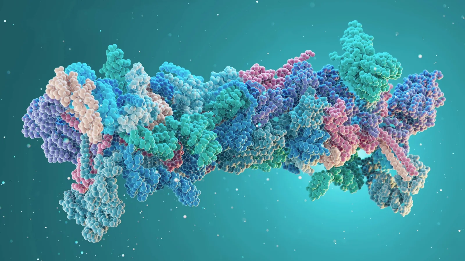
The α-helix is one of the two main secondary structures of proteins (along with the β-sheet). Described by Linus Pauling and Robert Corey in 1951, it is present in about 30% of residues in globular proteins and is the dominant structure in fibrous proteins such as keratin and myosin.
2. Structure of the α-Helix
The α-helix is a right-handed helix with 3.6 residues per turn and a pitch of 0.54 nm. Each amino acid is offset by 100° from the previous one. The peptide backbone forms the helix axis, with side chains pointing outward. Hydrogen bonds form between the C=O group of residue i and the N-H group of residue i+4, stabilizing the structure.
3. Helix Stabilization
The stability of the α-helix depends on the nature of the amino acids. Proline is a "helix breaker" because its pyrrolidine ring prevents formation of the required hydrogen bond. Same-sign charged amino acids repel each other and destabilize the helix. Glycine, too flexible, favors other conformations. Amphipathic helices have a hydrophobic face and a hydrophilic face, making them useful for membrane interactions.
4. Biological Functions
- Keratin: supercoiled α-helices forming hair, nails and horns
- Myosin: helical tail enabling assembly into thick filaments
- Hemoglobin: 8 α-helices per subunit framing the heme group
- Ion channels: transmembrane helices forming the pore
- Transcription factors: DNA-recognition helices (helix-turn-helix motif)
5. Conclusion
The α-helix is an optimal geometric solution for organizing a polypeptide chain: it maximizes intramolecular hydrogen bonds while allowing great functional variety depending on the nature of side chains. Its ubiquitous presence in proteins reflects the thermodynamic robustness of this structure.
The relation to the site’s thesis is precise: the alpha helix as a local compromise between geometry and bonding may share geometry, dynamics, or representation with another family of phenomena, but these levels are not identical. Comparison becomes scientific when it names the proposed shared mechanism, the variables that differ, and the test that could distinguish them.
The relation to the site’s thesis is precise: the alpha helix as a local compromise between geometry and bonding may share geometry, dynamics, or representation with another family of phenomena, but these levels are not identical. Comparison becomes scientific when it names the proposed shared mechanism, the variables that differ, and the test that could distinguish them.
The relation to the site’s thesis is precise: the alpha helix as a local compromise between geometry and bonding may share geometry, dynamics, or representation with another family of phenomena, but these levels are not identical. Comparison becomes scientific when it names the proposed shared mechanism, the variables that differ, and the test that could distinguish them.
The relation to the site’s thesis is precise: the alpha helix as a local compromise between geometry and bonding may share geometry, dynamics, or representation with another family of phenomena, but these levels are not identical. Comparison becomes scientific when it names the proposed shared mechanism, the variables that differ, and the test that could distinguish them.
The relation to the site’s thesis is precise: the alpha helix as a local compromise between geometry and bonding may share geometry, dynamics, or representation with another family of phenomena, but these levels are not identical. Comparison becomes scientific when it names the proposed shared mechanism, the variables that differ, and the test that could distinguish them.
Approaching the phenomenon without confusing image and mechanism
The concrete starting point is the alpha helix as a local compromise between geometry and bonding. An alpha helix is a secondary structure of a polypeptide chain. It results from regular geometry stabilized by hydrogen bonds between backbone groups. It is not a planar spiral: the chain advances along an axis and has chirality. Shape also depends on residues and environment. This first distinction matters because a winding form can be a trajectory, a structure, a projection, or simply a graphical convention.
The model that makes comparison testable
Dihedral angles φ and ψ describe local rotations around the backbone. Some combinations are favorable; others create steric clashes. The observed structure is therefore a solution in a constrained conformational space, not a mere aesthetic preference for a wound curve. [1] The model is not the object: it is a description whose assumptions must remain visible.
An example, then its counter-example
A Ramachandran plot represents accessible regions of these angles and helps distinguish helices, sheets, and unfavorable conformations. An experimental structure adds resolution, temperature, sequence, and molecular context. A helix icon is not enough to identify a real protein. The counter-example is as instructive as the example: it shows which variable is missing when moving from one domain to another.
What can actually be measured
A pedagogical figure can display axis, helical pitch, handedness, and hydrogen-bond positions. It should also show the chain as a sequence of residues, not as a chemistry-free continuous ribbon. This prevents helix, spiral, and macroscopic spring from being conflated. A quantitative figure must distinguish published data, theoretical calculation, simulation, and conceptual diagram.
Decomposing variables instead of following the silhouette
To understand the alpha helix as a local compromise between geometry and bonding, separate at least the variable that rotates, the one that changes scale, and the one that transports information or matter. An alpha helix is a secondary structure of a polypeptide chain. It results from regular geometry stabilized by hydrogen bonds between backbone groups. It is not a planar spiral: the chain advances along an axis and has chirality. Shape also depends on residues and environment. A global form can remain stable while its components are replaced; conversely, a trajectory can change shape without a new local mechanism.
The counter-example that protects the reasoning
Visual convergence comes from repeated orientation constraints. The real mechanism is bonding, sterics, and solvent physics. A molecular helix is therefore a case where shape resembles a spiral but the relevant dimension is three-dimensional and chemical. We therefore cannot conclude from an image alone. We must ask which observation would falsify the hypothesis, then keep negative results in the comparison. This discipline is especially important when several models produce a similar outline.
A reproducible method
A pedagogical figure can display axis, helical pitch, handedness, and hydrogen-bond positions. It should also show the chain as a sequence of residues, not as a chemistry-free continuous ribbon. This prevents helix, spiral, and macroscopic spring from being conflated. To make the result checkable, preserve parameters, units, resolution, initial conditions, and calculation method. An interactive visualization should also provide a readable initial state and a reset control; otherwise readers see an output without understanding its dependencies.
Open questions
The model explains dihedral angles φ and ψ describe local rotations around the backbone. some combinations are favorable; others create steric clashes. the observed structure is therefore a solution in a constrained conformational space, not a mere aesthetic preference for a wound curve. [1] but does not answer questions at every scale. We still need to determine how initial conditions, perturbations, and system boundaries alter the pattern. This is where research begins: not by adding an analogy, but by formulating a prediction that could be measured.
Limits and scope of the analogy
Visual convergence comes from repeated orientation constraints. The real mechanism is bonding, sterics, and solvent physics. A molecular helix is therefore a case where shape resembles a spiral but the relevant dimension is three-dimensional and chemical. [2]
Where the spiral is — and where it is not
The relation to the site’s thesis is precise: the alpha helix as a local compromise between geometry and bonding may share geometry, dynamics, or representation with another family of phenomena, but these levels are not identical. Comparison becomes scientific when it names the proposed shared mechanism, the variables that differ, and the test that could distinguish them.
References
- [1]Pauling, Linus et al.. (1951). The Structure of Proteins: Two Hydrogen-Bonded Helical Configurations of the Polypeptide Chain. Proceedings of the National Academy of Sciences. DOI: 10.1073/pnas.37.4.205
- [2]Ramachandran, G. N. et al.. (1963). Stereochemistry of Polypeptide Chain Configurations. Journal of Molecular Biology. DOI: 10.1016/S0022-2836(63)80023-2